

Retinoblastoma Diagnosis
Good vision is a sign of the correct comprehensive development of the child. The inability to see objects negatively affects the psychological state of the baby, oppresses him, prevents him from studying well at school, being safe on the street. And the task of parents is to help identify visual impairment in time for doctors who will conduct an examination and prescribe corrective treatment based on its results.
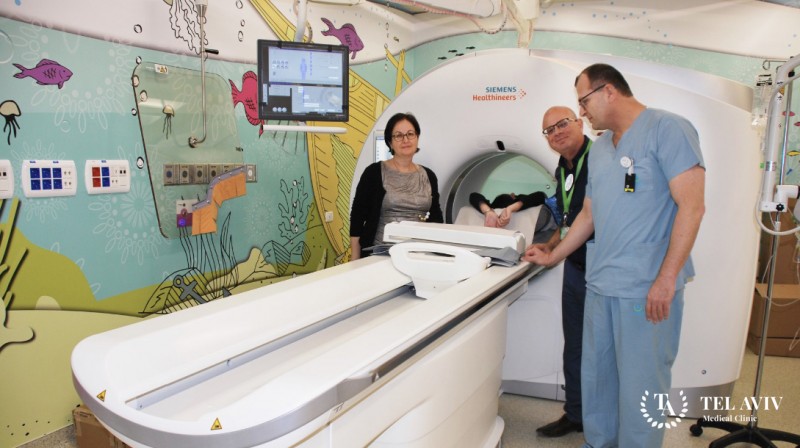
This requires special and modern equipment, which is available in the Israeli clinic Tel Aviv Medical Clinic. On the latest equipment, experienced children’s ophthalmologists, using innovative technologies, will determine the presence and absence of violations. And the high qualification of doctors of the pediatric department of hemato oncology allows us to achieve excellent results in eliminating pathology.
Symptoms of pathology
Retinoblastoma is a rare form of cancer, affecting the eye retina. And the disease occurs mainly in children under the age of 5. The tumor can affect both one eye and both. The treatment of such a pathology is a rather complex and long-lasting process. Therefore, the child and his parents should stock up on patience, increase their strength to combat a dangerous disease.
The sooner parents suspect the problem, the faster doctors can get rid of it. But the insidiousness of oncology in its asymptomatic course at the initial stages of development. With the growth of education, the following signs appear:
- Discoloration of the pupil.
- Visual impairment.
- Beginning strabismus.
- Pain in the eye.
- Retinal redness.
- Swelling of the eye.
Diagnosis of pathology
Vision at the Tel Aviv Medical Clinic is checked on ultramodern devices that do not cause unpleasant feelings or discomfort in the child. Our experts know how to properly interact with a small patient so that he does not whimper, but allows him to calmly check his eyes. We use safe contactless research techniques on sensitive devices, which together gives 100% diagnostic accuracy.
- Ocular floor imaging is done by using a binocular ophthalmoscope.
- Corneal parameters and refraction of light in the optical structure of the eye is determined by an autorefractometer.
- Visual acuity is assessed by a phoropter with a built-in computer system.
- Intraocular pressure is checked by pneumotonomer.
- Field of view measures the perimeter method.