


Anatomy of the Eye
Retinoblastoma is a rare type of eye cancer. The tumour grows in the retina of the eye (the medical term for retina is retina) and is almost always found only in children. This disease has a hereditary and non-hereditary form. If retinoblastoma is hereditary, then family members have a predisposition to the fact that one of them may get sick. If retinoblastoma is not hereditary, then eye cancer occurs spontaneously, that is, the cells of the retina of the eye begin to change by themselves. Retinoblastoma can affect one eye or both eyes. Most often, it grows in only one eye. Retinoblastomas usually grow quickly. They can appear inside the eyeball.
The leading Israeli Tel Aviv Medical Clinic employs experienced doctors who have been treating eye diseases in children for more than 10 years. We use the latest equipment and unique methods of therapy. Our employees annually undergo internships in foreign clinics. It is worth noting that we have set tariffs at the state level. Thus, you receive high-quality services for which you pay less.
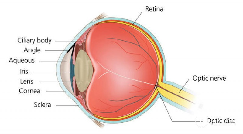
Anatomy of the eye
The human eye is spherical, hence its name is an eyeball. It consists of three shells: the external, vascular and retinal, as well as internal contents. The front of the outer shell – the cornea – is like a transparent window into the outside world, through it rays of light enter the eye. Having a convex shape, it not only passes but also refracts these rays. The rest of the outer shell – the sclera – is opaque and looks like boiled egg white.
The second shell – vascular – consists of many small vessels, through which blood supplies the eye with oxygen and nutrients. Several parts are also distinguished in this shell: the anterior – the iris, the middle – the ciliary body and the posterior – the choroid. In the centre of the iris is a round hole – a pupil. The space between the cornea and the iris is called the anterior chamber. The ciliary body produces intraocular fluid that circulates inside the eye, washing and feeding on the cornea, lens, vitreous body. Choroid – the posterior part of the vascular membrane – directly contacts the retina, providing it with the necessary nutrition.
The third sheath of the eye – the retina (or retina) – consists of several layers of nerve cells and lines it from the inside. It is she who provides us with a vision. The retina displays the objects we see. Inside the shells, there are anterior and posterior (between the iris and lens) chambers filled with ocular fluid inside, and most importantly, the lens and vitreous body.